
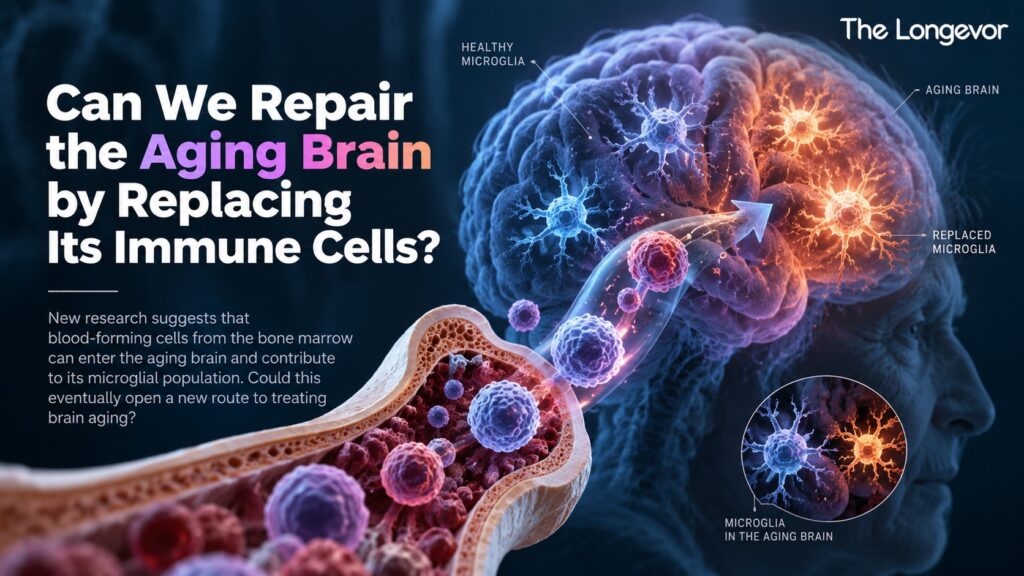
Can We Repair the Aging Brain by Replacing Its Immune Cells?
New human research challenges a long-standing view of brain immunity, revealing that blood-forming cells from the bone marrow can enter the aging brain and contribute to its microglial population. Could this eventually open a new route to treating brain aging?
For decades, scientists have viewed the brain’s immune system as unusually self-contained. At the center of that system are microglia — specialized immune cells that live within the brain and help maintain its environment. They remove cellular debris, respond to injury, interact with neurons and help regulate the brain’s immune response.
Unlike many immune cells in the body, microglia were thought to be largely established early in development and maintained locally throughout life. New research in humans is challenging that assumption.
A study published in Nature in 2026 found evidence that bone-marrow-derived cells enter the human brain during aging and can contribute substantially to the population of cells resembling microglia. The researchers detected this phenomenon in all 20 older individuals examined.
The finding does not mean that the brain simply replaces its old immune cells with new ones as we age. Nor does it demonstrate that replacing microglia can reverse cognitive aging. But it does reveal something potentially important: the aging human brain may be more connected to the body’s circulating immune system than scientists previously thought.
And that could change how researchers think about treating age-related brain disorders.
What Are Microglia?
Microglia are often described as the resident immune cells of the brain. They belong to the broader family of macrophage-like immune cells and perform several functions that are essential for maintaining brain health.
They monitor their surroundings, clear cellular waste, respond to injury and infection, and communicate with neurons and other cells. Microglia also participate in the remodeling of neural connections. Under healthy conditions, these activities help maintain the brain’s internal environment.
But aging changes microglia. Older brains contain microglial populations with altered gene-expression patterns and inflammatory states. Some microglia can become more reactive, while others adopt disease-associated or senescence-related characteristics.
Recent research has increasingly linked these changes to the biology of neuroinflammation and age-related brain dysfunction. This has raised a fundamental question: If aging changes the brain’s resident immune cells, can those cells ever be replaced?
Until recently, the answer appeared to be largely “no” — at least under normal conditions.
The Long-Standing Microglia Model
Much of what scientists know about microglial development comes from mouse research. In mice, microglia enter the brain during embryonic development and can maintain themselves through local self-renewal for most of the animal’s life.
Adult blood-forming cells contribute relatively little to the normal microglial population under healthy conditions. This created a simple model:
Microglia are born early → enter the brain → remain there → renew themselves locally.
But humans may not follow exactly the same biological rules. The new Nature study set out to investigate that question directly.
Scientists Found Evidence of Blood-Derived Cells in the Aging Human Brain
Researchers developed a method that uses somatic mutations — genetic changes that accumulate naturally in cells over a person’s lifetime — as biological markers of cell ancestry.
The idea is surprisingly powerful. Cells that originate from the same developmental lineage can share patterns of accumulated mutations. By studying these mutations, researchers can reconstruct relationships between cells and determine where particular populations came from.
The team applied this approach to brain tissue from 20 older individuals. They found evidence of an influx of bone-marrow-derived cells into the brain in every individual examined.
Single-cell analyses, including lineage tracing using mitochondrial DNA variants, showed that these infiltrating cells could closely resemble microglia and, in some individuals, constitute a substantial fraction of the microglial population.
That is the key discovery: the aging human brain is not necessarily an entirely closed immune environment. Cells originating from outside the brain can become part of its resident-like immune-cell population.
Why This Is Different From What Scientists Expected
The finding is particularly interesting because the blood-brain barrier is one of the body’s major protective interfaces. The barrier helps control which cells and molecules can move between the bloodstream and the brain.
Most circulating immune cells do not simply enter healthy brain tissue whenever they want. Yet the researchers found evidence that bone-marrow-derived cells become increasingly represented within the human brain’s microglial population with age.
This doesn’t necessarily mean that the blood-brain barrier becomes broadly “leaky.” Instead, the findings suggest that the relationship between the peripheral immune system and the aging brain is more dynamic than previously appreciated.
That raises an important possibility: Could changes in the body’s immune system influence the immune environment of the brain during aging?
Does This Mean Old Microglia Are Being Replaced?
Not exactly. This distinction is critical.
The study provides evidence that bone-marrow-derived cells contribute to the microglial pool, but it does not show that all existing microglia disappear and are replaced by younger cells. The brain still contains its original resident microglia.
Instead, the research suggests that another population of myeloid cells can enter the brain and acquire characteristics similar to microglia. In other words, aging may create a more mixed immune-cell landscape inside the human brain.
That is very different from saying:
“The brain replaces all its old immune cells.”
The actual biology is more nuanced.
Why Would This Matter for Brain Aging?
One reason the discovery matters is that microglia change significantly during aging. Aged microglia can become less efficient at maintaining tissue homeostasis and can adopt inflammatory states.
Persistent neuroinflammation is increasingly recognized as one component of brain aging and neurodegenerative disease biology. If bone-marrow-derived cells are entering the brain more frequently with age, researchers now have to ask: What determines whether these incoming cells are beneficial, harmful or simply different?
Their behavior may depend on their developmental origin, the environment they encounter inside the brain and the signals they receive from aging neural tissue.
The new study does not yet answer that question, but it makes the question experimentally accessible.
The Alzheimer’s Connection Is Particularly Interesting
The researchers also examined human cohort data involving clonal hematopoiesis, a phenomenon in which blood-forming stem or progenitor cells acquire mutations and expand into larger cellular clones.
They found an association between most types of clonal hematopoiesis examined and a lower prevalence of Alzheimer’s disease-related outcomes in their analysis. That observation is intriguing because it suggests that certain blood-cell populations may have effects within the aging brain that are not necessarily harmful.
However, it is important not to overinterpret this result. The study demonstrates an association, not proof that clonal hematopoiesis protects people from Alzheimer’s disease.
It does not mean that acquiring blood-cell mutations is beneficial, nor does it suggest that anyone should attempt to alter their blood-cell populations. The finding instead provides another clue that peripheral immune-cell biology and brain aging may be connected.
Could We Use Blood-Born Cells to Repair the Aging Brain?
This is where the research becomes especially interesting for longevity science.
If bone-marrow-derived cells can naturally enter the aging brain, scientists may eventually be able to ask whether these cells could be engineered or selected for therapeutic purposes.
Imagine, in principle, a cell originating outside the brain that could enter the central nervous system and perform a specific function. It might potentially:
- Deliver a therapeutic protein
- Replace a defective immune-cell function
- Reduce harmful inflammatory signaling
- Support damaged neural tissue
- Correct a disease-associated cellular pathway
This concept is not entirely theoretical. Researchers have already been studying microglia replacement and transplantation strategies for certain neurological diseases and genetic disorders. Experimental work has shown that replacing dysfunctional microglia can have therapeutic effects in specific disease models.
But these approaches are still highly experimental.
The new human study does something different. Rather than demonstrating a therapy, it provides evidence that the human brain has a natural route through which blood-derived cells can become part of its immune-cell landscape during aging.
That could potentially make future cell-based strategies more feasible.
But Can Replacing Microglia Actually Make the Brain Younger?
We don’t know. And this is where the distinction between biological discovery and therapeutic promise matters.
The study does not show that the incoming cells are younger, healthier or more functional than the microglia they join. It also does not show that people with more bone-marrow-derived microglia have better memory, slower cognitive decline or lower risk of dementia.
Most importantly, researchers have not demonstrated that deliberately increasing this process improves brain health in aging humans.
The study is therefore better understood as a map of previously underappreciated biology, rather than a treatment breakthrough.
The Identity of a Microglial Cell May Matter Less Than Its Environment
One of the deeper implications of this work is that cell identity may be more flexible than previously assumed. Microglia are shaped not only by their origin but also by the environment in which they live.
The researchers found that infiltrating bone-marrow-derived cells can become similar to microglia at the molecular level once inside the brain. This supports a broader idea in modern aging research:
The tissue environment can influence what a cell becomes and how it behaves.
That has important implications for longevity. If an aging brain creates an inflammatory or dysfunctional environment, simply introducing new cells may not be enough. Those cells would still encounter the same signals.
So a future therapy might need to address both the cells themselves and the environment surrounding them.
Why This Could Matter Beyond Microglia
The discovery also fits into a larger shift in how researchers understand aging. The brain is not an isolated organ. It communicates continuously with the immune system, blood vessels, endocrine system and peripheral organs.
Immune cells circulate through the body, metabolites travel through the bloodstream, and inflammatory signals can influence distant tissues. Aging changes all of these systems simultaneously.
Recent research increasingly supports the idea that brain aging is a systems-level process, involving interactions among neurons, glial cells, immune populations, blood vessels and metabolic pathways.
The new Nature study adds another piece to that picture. The immune cells inside an aging brain may themselves be partly connected to the body’s blood-forming system.
What We Still Don’t Know
Several major questions remain.
Are the incoming cells beneficial or harmful?
The study shows that bone-marrow-derived cells can contribute to the microglial pool, but it does not establish that these cells are universally beneficial. Their effects may vary depending on their molecular state and the environment of the aging brain.
Why does this happen more frequently with age?
The precise mechanisms controlling this increased influx remain an important area for future research. Changes in the blood-brain barrier, brain inflammation, vascular biology or immune signaling could potentially contribute, but the current study does not establish one simple cause.
Do these cells improve brain function?
There is currently no evidence from this study that greater replacement leads to better memory or slower cognitive decline. That question requires functional studies.
Could these cells be engineered?
Potentially, this is an exciting research direction. But engineered immune-cell therapies for the brain would require extensive testing for safety, targeting, persistence and unintended immune effects.
Could this help treat Alzheimer’s disease?
It is far too early to say. The association with Alzheimer’s-related outcomes is intriguing, but it does not prove that changing microglial origin or replacement would prevent or treat Alzheimer’s disease. Clinical trials would ultimately be required.
The Bigger Question: Can the Aging Brain Be Rebuilt?
For years, the idea of repairing an aging brain sounded almost impossible. Neurons are highly specialized, the blood-brain barrier limits access, and microglia were considered a largely self-maintaining population established early in life.
The new findings don’t solve those problems. But they change one important assumption: the aging human brain may not be as immunologically closed as scientists once believed.
If blood-derived cells can enter the brain and become part of its microglial population, then the peripheral immune system may offer an unexpected route for influencing brain biology.
That does not mean we can currently replace old microglia and reverse brain aging. But it suggests that researchers may have more biological options than previously thought.
The Longevor Takeaway
A new Nature study has challenged a long-standing view of how the human brain maintains its immune cells. By tracking naturally occurring somatic mutations and using single-cell analyses, researchers found evidence that bone-marrow-derived cells enter the aging human brain and can make up a substantial fraction of the microglial population. The phenomenon was observed across all 20 older individuals examined.
The finding is important because it reveals a previously underappreciated connection between the peripheral immune system and the aging brain.
But the next step is not to assume that replacing microglia will make the brain younger. Researchers first need to determine which incoming cells are beneficial, how they behave inside the aging brain, why their numbers increase with age and whether manipulating this process can improve brain function.
For longevity science, the most interesting possibility is not simply replacing an old cell with a new one. It is understanding whether the immune environment of the aging brain can be deliberately reshaped.
If future research can answer that question, cells produced outside the brain could eventually become part of a new generation of therapies designed to influence how the brain ages.
For now, however, the discovery represents something more fundamental:
The aging brain may be more connected to the body’s immune system than we once believed.